
Understanding the structural organization in animals is fundamental to comprehending how various bodily functions are carried out. This encompasses the study of tissues, which are groups of similar cells performing specific functions. The primary types of animal tissues include epithelial, connective, muscular, and nervous tissues. Below is a detailed exploration of these tissues and their subtypes.
1. Epithelial Tissue

Epithelial tissue forms the covering or lining of all internal and external body surfaces. The cells in epithelial tissues are closely packed, with minimal intercellular space, and are bound together by specialized junctions. This tissue serves functions such as protection, absorption, secretion, and sensation.
a. Simple Epithelium
Simple epithelium consists of a single layer of cells and is primarily involved in processes like absorption, secretion, and filtration. Based on cell shape, simple epithelium is classified into:

- Simple Squamous Epithelium: Composed of flat, thin cells with irregular outlines, this type facilitates diffusion and filtration. It lines structures such as alveoli in the lungs and blood vessels.
- Simple Cuboidal Epithelium: Made up of cube-shaped cells, it functions in secretion and absorption. This epithelium is found in glands and their ducts, as well as in kidney tubules.
- Simple Columnar Epithelium: Consisting of tall, column-like cells, it is specialized for absorption and secretion. This type lines the stomach, intestines, and parts of the respiratory tract.
- Ciliated Epithelium: A subtype where cells possess cilia on their free surface, aiding in the movement of substances. It is present in the respiratory tract and fallopian tubes.
b. Compound (Stratified) Epithelium
Compound epithelium comprises multiple layers of cells, providing protection against mechanical and chemical stresses. The classification includes:

- Stratified Squamous Epithelium: The outermost layer consists of flat cells. It can be keratinized (as in the skin) or non-keratinized (as in the oral cavity and esophagus).
- Stratified Cuboidal Epithelium: Typically two layers of cube-shaped cells, found in ducts of sweat glands and mammary glands.
- Stratified Columnar Epithelium: Surface cells are columnar, while basal layers are cuboidal. This type is rare but can be found in parts of the male urethra and some glandular ducts.
c. Cell Junctions
Cell junctions are specialized structures that connect adjacent cells, facilitating communication and maintaining tissue integrity. The main types include:

- Tight Junctions (Occluding Junctions): Seal adjacent cells to prevent the passage of molecules between them, maintaining distinct compartments within tissues.
- Adherens Junctions: Connect the actin cytoskeleton of one cell to another, providing mechanical stability.
- Desmosomes: Bind intermediate filaments of adjacent cells, offering resistance to mechanical stress.
- Gap Junctions: Form channels that allow direct communication between the cytoplasm of adjacent cells, enabling the passage of ions and small molecules.
2. Connective Tissue
Connective tissue supports, binds, and protects other tissues and organs. It is characterized by an abundance of extracellular matrix, which determines its properties. The primary components include:
- Cells: Such as fibroblasts (produce fibers and ground substance), adipocytes (store fat), macrophages (phagocytic cells), and mast cells (involved in inflammatory responses).
- Fibers: Including collagen fibers (provide strength and flexibility), elastic fibers (allow stretch and recoil), and reticular fibers (form supportive networks).
- Ground Substance: An amorphous material filling the space between cells and fibers, composed of proteoglycans and glycosaminoglycans, which retain water and contribute to the tissue’s resilience.
Connective tissue is classified into various types:
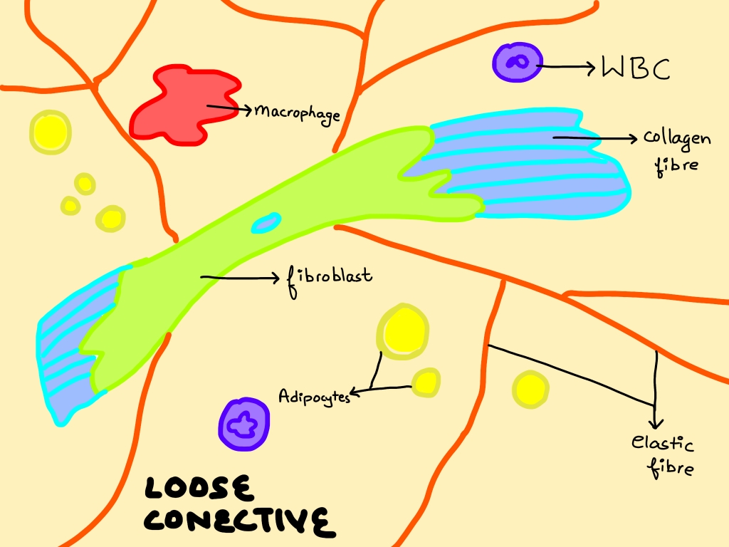
a. Loose Connective Tissue
- Areolar Tissue: A loose arrangement of fibers and cells, providing support and flexibility. It underlies epithelia and surrounds blood vessels and nerves.
- Adipose Tissue: Specialized for fat storage, it insulates the body and cushions organs.
- Reticular Tissue: Contains a network of reticular fibers supporting soft tissues like the liver, spleen, and lymph nodes.

b. Dense Connective Tissue
- Dense Regular Connective Tissue: Collagen fibers are aligned in parallel, providing tensile strength in one direction. Tendons and ligaments are examples.
- Dense Irregular Connective Tissue: Collagen fibers are arranged irregularly, offering strength in multiple directions. It is found in the dermis of the skin and organ capsules.
c. Specialized Connective Tissue
- Cartilage: A firm yet flexible tissue lacking blood vessels. Chondrocytes (cartilage cells) are embedded in a matrix rich in collagen and proteoglycans. Types include:
- Hyaline Cartilage: Provides support with some flexibility; found in the nose, trachea, and at the ends of long bones.
- Elastic Cartilage: Contains elastic fibers, allowing more flexibility; found in the external ear and epiglottis.
- Fibrocartilage: Rich in collagen fibers, it is extremely durable and tough; found in intervertebral discs and the menisci of knee joints.
- Bone: A rigid tissue providing structural support. Osteocytes (bone cells) are embedded in a mineralized matrix containing collagen fibers and inorganic salts like calcium phosphate. Bone is classified into:
- Compact Bone: Dense and organized into structural units called osteons (Haversian systems). Found in the outer layers of bones, it provides strength and support.
- Spongy Bone (Cancellous Bone): Contains a porous, trabecular structure, making it lighter. It houses bone marrow, which produces blood cells. Found at the ends of long bones.
Structure of a Long Bone
A long bone, such as the femur or humerus, has the following parts:

- Diaphysis: The long, cylindrical shaft composed of compact bone surrounding the medullary cavity (marrow cavity).
- Epiphysis: The enlarged ends of the bone, consisting mainly of spongy bone covered by a thin layer of compact bone.
- Metaphysis: The region between the diaphysis and epiphysis, containing the epiphyseal plate (growth plate) in growing bones.
- Periosteum: A fibrous membrane covering the outer surface of bones, containing blood vessels and nerves for bone nourishment and repair.
- Endosteum: A thin membrane lining the medullary cavity, housing bone-forming cells.
- Bone Marrow: Present inside the medullary cavity; red marrow is involved in blood cell production, while yellow marrow stores fat.
3. Muscular Tissue
Muscular tissue consists of elongated cells called muscle fibers, which are responsible for movement. Muscle cells contain contractile proteins, actin, and myosin.
Types of Muscular Tissue

- Skeletal Muscle:
- Structure: Long, cylindrical, multinucleated fibers with striations.
- Function: Voluntary movement; attached to bones and responsible for locomotion.
- Location: Biceps, triceps, diaphragm, etc.
- Smooth Muscle:
- Structure: Spindle-shaped, uninucleated, non-striated fibers.
- Function: Involuntary control; regulates functions like digestion and blood vessel constriction.
- Location: Walls of hollow organs (intestines, blood vessels, uterus).
- Cardiac Muscle:
- Structure: Branched, uninucleated fibers with striations and intercalated discs.
- Function: Involuntary contraction; ensures rhythmic heartbeats.
- Location: Heart walls.
4. Nervous Tissue
Nervous tissue is specialized for transmitting electrical impulses, coordinating and controlling bodily functions. It comprises two main components:

a. Neuron (Nerve Cell)
- Structure:
- Cell Body (Soma): Contains the nucleus and cytoplasmic organelles.
- Dendrites: Short, branched projections that receive impulses from other neurons.
- Axon: A long, single fiber that transmits impulses away from the cell body. Some axons are covered by a myelin sheath, which speeds up signal transmission.
- Function: Neurons conduct electrical signals between different parts of the body, enabling sensation, movement, and cognition.
b. Neuroglial Cells (Glial Cells)
- Function: Support, protect, and nourish neurons. They do not conduct nerve impulses.
- Types:
- Astrocytes: Maintain the blood-brain barrier and support neurons.
- Oligodendrocytes: Form the myelin sheath in the central nervous system (CNS).
- Schwann Cells: Produce myelin in the peripheral nervous system (PNS).
- Microglia: Act as immune cells in the nervous system, removing pathogens and debris.